Magnetic Resonance Imaging (MRI)
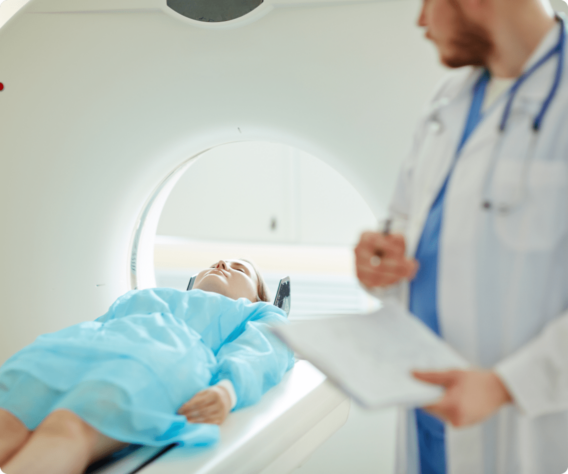
Magnetic Resonance Imaging (MRI) provides a remarkable window into the body’s internal structures. It has become an invaluable diagnostic tool, replacing many invasive surgical procedures.
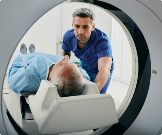
Unlike other techniques, MRI does not use radiation. It combines a powerful magnetic field, radio frequency pulses, and a computer to generate highly detailed images of soft tissue structures near and around bones, blood vessels, organs, and the brain.
How MRI Works
The human body is mostly water, which contains hydrogen. The MRI scanner’s magnetic field causes hydrogen protons to align and spin in the same direction. The machine then emits a radio frequency pulse. This pulse causes the protons to absorb energy and spin in a slightly different direction. As the protons gradually release this absorbed energy, the machine measures the energy and uses a computer to mathematically construct detailed images.
MRI gives us minutely thin, cross-sectional views or “slices” of anatomic structure, revealing even the most subtle differences in body tissues. It can be tailored to answer specific diagnostic questions.
Preparing for Your MRI
An MRI exam generally requires minimal preparation. For many MRI exams, you can eat, drink, and take your medications as normal.
However, some kinds of MRI exams require you to abstain from all eating and drinking for two to four hours beforehand. Ask your doctor’s office about any special preparations you may need.
If you have any questions, please check with your doctor’s office, or phone one of our imaging technologists at (541) 269-8090.
