Advanced Diagnostic Technology

The high-tech marvels of today’s medical imaging provide detailed information to target your diseases as never before. Performing more than 60,000 imaging procedures each year, Bay Area Hospital is the premier imaging site in southwestern Oregon.

Our highly trained, friendly staff of imaging professionals provides exceptional care for our patients. A team of board certified radiologists will interpret your test results and provide expert support to your physician.

Our goal is to provide the highest quality of diagnostic imaging, serving every patient with professionalism, compassion, and integrity.

Computer Tomography

Fast, Safe, Comfortable

A CT scan combines multiple X-ray images from different angles to produce cross-section views of the body. Our advanced CT device, the Aquilion PRIME, can produce 160 of these virtual “slices” per rotation. It rapidly yields exceptional images while exposing the patient to far less radiation than less advanced devices. With a wider couch and gantry than most CTs, it also ensures patient comfort and convenience.

Computed tomography (CT) uses an X-ray source and a computer to create multiple CT images. The final pictures are a far more detailed diagnostic tool than a single X-ray film.

For patients, the CT experience is quick, easy, and comfortable. The machine looks like a doughnut-shaped ring, not a tunnel. Typically, the only pain you feel is the poke of a needle to deliver a contrast material into your bloodstream. If no contrast is required, there will be no pain at all.

Inside the CT scanner is an X-ray detector, which can distinguish hundreds of levels of density. With the help of swallowed and injected contrast material, the scanner can see the different tissues inside any solid organ.

During the scan, the X-ray beam makes a spiral path. Scanning in a helical motion (like a Slinky), it gathers continuous data with no gaps between the images. This data is transmitted to a computer, which builds and displays 2-D and 3-D cross-sectional pictures of any part of the body.

To make CT exams as fast, safe and accurate as possible, Bay Area Hospital employs the Aquilion PRIME CT system from Toshiba America Medical Systems Inc. Aquilion PRIME provides faster exams and produces high-quality images required for accurate diagnoses. Yet its X-ray doses are as low as reasonably achievable, thanks to industry-leading dose-reduction software.

The speed of the Aquilion PRIME enables our clinicians to obtain critical patient information for enhanced diagnoses faster than ever before. The Aquilion PRIME’s large bore opening offers a more spacious feeling for patients, while a higher table weight limit accommodates a full range of patients.

This advanced device represents Bay Area Hospital’s commitment to leading-edge technology, providing excellent diagnostic information to our doctors while assuring safety and comfort for our patients.

Different CT scans require different kinds of preparation, and some scans need no preparation at all. Be sure to ask your doctor’s office what preparation you need for your particular scan.

For most scans of your torso, you’ll be asked not to eat solid foods for four hours before the exam. During this time you should drink only clear liquids, such as black coffee, apple juice, broth, or water.

For some kinds of exams, you may be asked to drink a specified amount of water for a certain period of time before your exam. Some abdominal scans require you to drink contrast fluid, either one hour or four hours before your exam.

Other preparations:

  • Do not wear metal items such as zippers and buttons.
  • Let our staff know what medications you are taking.
  • Let our staff know if you could be pregnant, are breastfeeding, are allergic to contrast medium (dye), or are diabetic.

If you have any questions, please check with your doctor’s office, or phone one of our imaging technologists at (541) 269-8090.

When you come to Bay Area Hospital for a CT scan, you’ll check-in at our Digital Imaging desk. The friendly, professional staff will verify your laboratory tests and double-check your allergies and other health history. In some cases, you’ll be asked to drink contrast material that will enhance the CT images.

Next, you’ll meet your CT technologist, who will take you to the scan room. You will be asked to remove all jewelry and other metal objects. Depending on what part of your body is being scanned, you may need to partially disrobe. You’ll be asked to lie on a  table that passes through the bore of the gantry — the “doughnut hole.”

The technologist will give you a full explanation of the process. You probably will be given contrast material by intravenous injection, in addition to the material you already swallowed.

When the contrast material is administered, you may feel unusual sensations such as a “hot flash” or a metallic taste. These will pass quickly. The entire scan takes between five and 20 minutes, though a few specialized scans can last longer.

After your scan, the results will be reviewed by one of our board-certified radiologists, who will relay the information to your physician. We will recommend that you drink water to help flush the contrast material from your body. You shouldn’t see or feel any aftereffects.

In addition to bachelor’s degrees and radiography licenses, all of our CT technologists have advanced certification through the American Registry of Radiologic Technicians specifically for CT. They have an average of more than 10 years’ experience.

Medical Imaging

Digital Mammography

Relaxed, Respectful, Reassuring

Caring for you is the core of what we do, and your welfare is our top priority. Our skilled, compassionate staff uses the most up to date equipment to make your mammogram as comfortable and stress-free as possible.

A digital mammogram is a low-radiation procedure, providing state-of-the-art diagnostic images. Because the technology is digital – like a digital camera – there’s no film to develop. Your images will be available for immediate review by our highly qualified radiologists.

A digital mammogram typically requires almost no preparation. Generally, you only need to make sure you’re not wearing deodorants, perfumes, powders, or bath oils. But be sure to ask your doctor’s office about any special preparations that you may need.

A stereotactic biopsy requires extra preparation. If you’re undergoing this procedure, you should avoid aspirin and anticoagulants for seven days before the procedure. Don’t eat or drink anything for two hours before the procedure. Wear comfortable clothing and a bra.

If you have any questions, please check with your doctor’s office, or phone our imaging department at (541) 269-8090.

When you arrive for testing at Bay Area Hospital, you’ll be asked to change into a hospital gown, and a radiology technologist will ask about your health history. Feel free to ask questions of your own. Our unhurried schedule provides ample time for each patient to have a relaxed, respectful experience.

Your exam generally involves making two images of each breast. A soft, warm cushion known as a MammoPad provides a gentler, more pleasant experience and helps obtain an image showing more of the breast.

After your exam, you’ll get dressed and meet with the radiologist, who interprets or “reads” the images. Depending on what the radiologist sees, you may be scheduled for additional tests.

Our facility is accredited by the American College of Radiology. Our staff is credentialed by the American Registry of Radiologic Technologists, with advanced certification in mammography. In addition to standard mammography, we offer 3D mammography, advanced diagnostic imaging, such as ultrasound, stereotactic breast biopsy, and ductogram.

Mammogram

Magnetic Vision

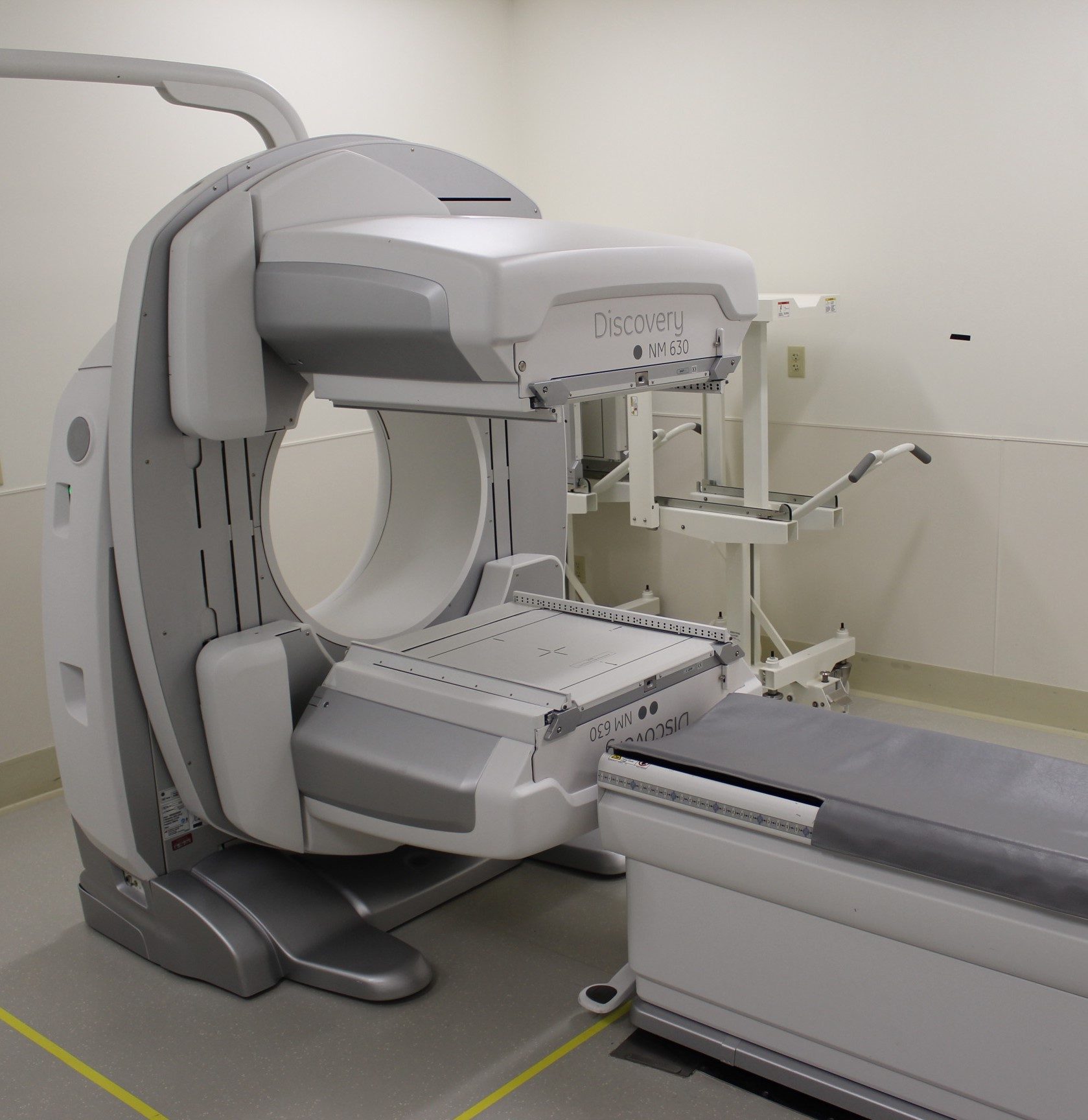
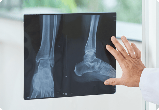
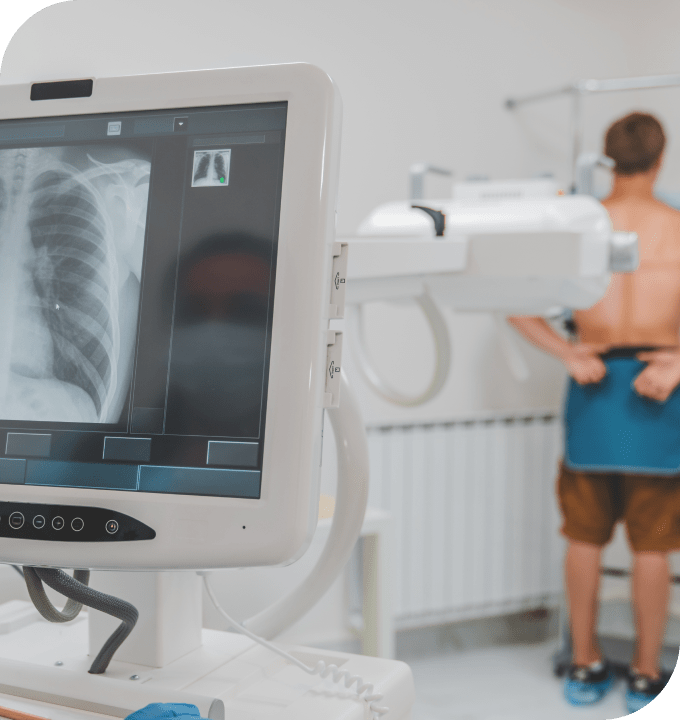
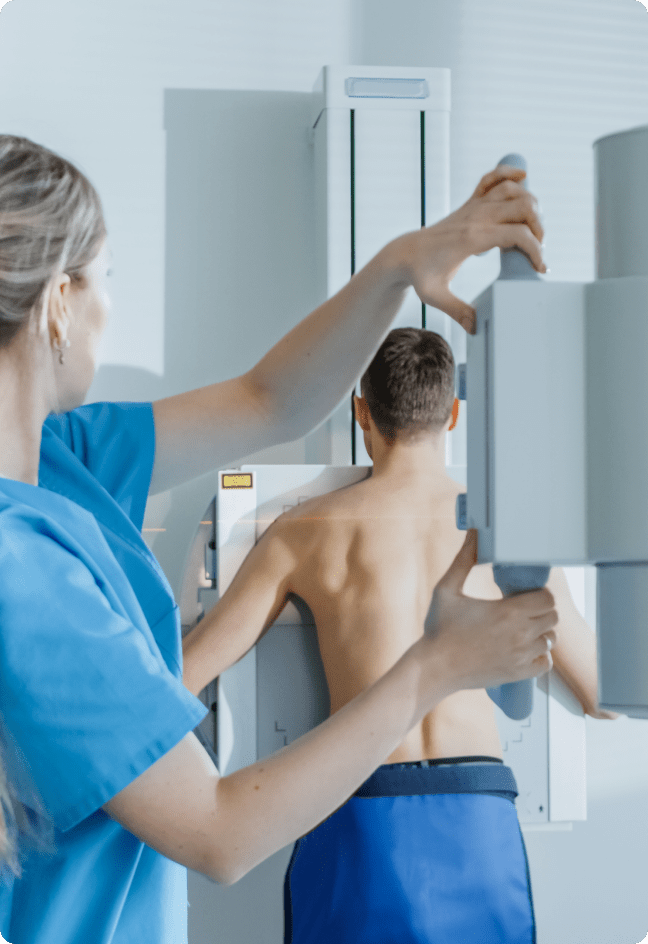
Magnetic Resonance Imaging (MRI) provides a remarkable window into the body’s internal structures. It has become an invaluable diagnostic tool, replacing many invasive surgical procedures.

Unlike other techniques, MRI does not use radiation. It combines a powerful magnetic field, radio frequency pulses, and a computer to generate highly detailed images of soft tissue structures near and around bones, blood vessels, organs and the brain.

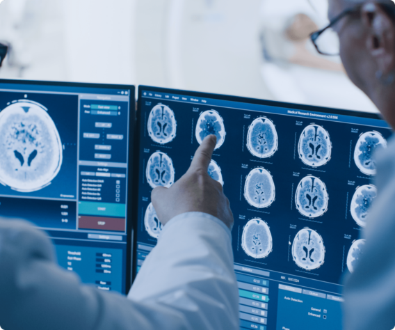
The human body is mostly water, which contains hydrogen. The MRI scanner’s magnetic field causes hydrogen protons to align and spin in the same direction. The machine then emits a radio frequency pulse. This pulse causes the protons to absorb energy and spin in a slightly different direction. As the protons gradually release this absorbed energy, the machine measures the energy and uses a computer to mathematically construct detailed images.

MRI gives us minutely thin, cross-sectional views or “slices” of anatomic structure, revealing even the most subtle differences in body tissues. It can be tailored to answer specific diagnostic questions.
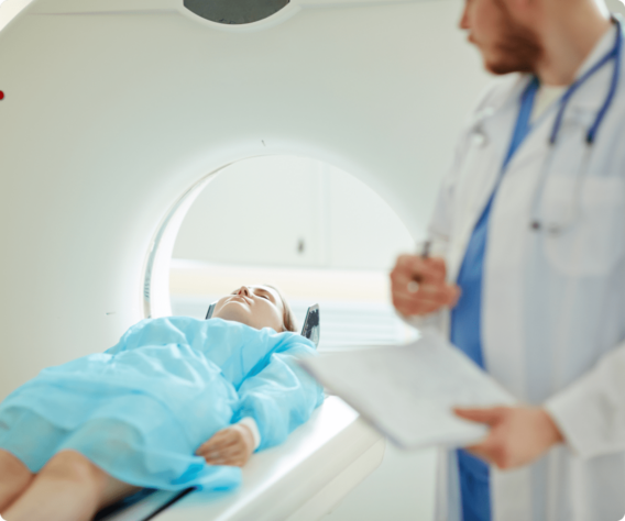
An MRI exam generally requires minimal preparation. For many MRI exams, you can eat, drink, and take your medications as normal.

However, some kinds of MRI exams require you to abstain from all eating and drinking for two to four hours beforehand. Ask your doctor’s office about any special preparations you may need.

If you have any questions, please check with your doctor’s office, or phone one of our imaging technologists at (541) 269-8090.

Because the MRI employs a powerful magnet, the presence of metal in your body may create a safety hazard. Before your MRI begins, tell the technologist if you have any metal or electronic devices in your body, such as an artificial heart valve, a metallic joint implant, a pacemaker, or shrapnel from an old wound.

You will be asked to change into a gown and to remove your watch, jewelry, dentures, eyeglasses, or other metal objects. Any metal in the room will react to the powerful magnet within the MRI machine.

For the same reason, do not take any credit, bank, parking, or insurance cards into the scan room. The magnet will erase the information on their magnetic strips.

During the procedure, you will hear loud clicking or tapping noises. These noises are produced when coils inside the MRI magnet are rapidly switched on and off. You’ll be given earplugs to muffle these sounds.

Your MRI procedure typically will take between 15 and 30 minutes. Patients generally find the experience comfortable and non-stressful.

Afterward, one of our board-certified radiologists will analyze your scan and report results to your doctor. Your doctor will discuss the findings with you.

The key difference among MRI scanners is the strength of their magnets, measured in units called teslas. The stronger the tesla strength, the better the image and the faster the exam.

Our top-of-the-line Magnetom Skyra MRI system has a superior magnetic field strength of 3 tesla. Large and roomy, with a short bore, it offers better patient comfort. No long-tunnel scanners are in use at our hospital.

Our MRI technologists have college degrees in radiology sciences, with specialized training in magnetic resonance imaging, and an average of more than 10 years’ experience. They are licensed by the Oregon Board of Medical Imaging and certified through the American Registry of Radiologic Technologists. Their proven expertise assures you safe and accurate imaging.

Patient And Medical Technician At The Computer Tomography

PET-CT

One Machine, Two Technologies

Combining two kinds of scans makes PET-CT a powerful diagnostic tool. It can pinpoint a tumor’s location, determine whether the cancer has spread, and measure how active the cancer is.

Combining the images of a positron emission tomography (PET) scan and a computed tomography (CT) scan, a PET-CT scan is a powerful diagnostic tool.

The CT scan provides detailed anatomical pictures of tissues and organs inside the body, while the PET scan reveals abnormally functioning tissues that can indicate cancer.

For a PET scan, glucose labeled with mildly radioactive tracers is injected into the patient. Cancerous tissue tends to attract this substance, which emits gamma rays that will be detected by the scanner. The PET scanner records these gamma rays from the various organs targeted for the exam.

In the CT portion of the procedure, a computer reassembles the signals into three-dimensional pictures of the body. These detailed, cross-sectional images show any abnormalities or tumors.

Combining these two kinds of scans creates a more complete image than either test alone. It can pinpoint a tumor’s location, determine whether the cancer has spread, and measure how active the cancer is.

PET-CT testing at Bay Area Hospital is conducted with a mobile scanner mounted on a trailer. Circulating among three Oregon hospitals, the mobile unit serves patients in Coos Bay one or two days each week, delivering the benefits of advanced technology with the convenience of close-to-home accessibility.

Do not eat or drink anything for six hours before the scan. This fasting will lower the overall sugar level in your body, making the glucose injection more visible.

You’ll need to be relaxed for the exam. So wear loose-fitting clothes and avoid caffeine. If you are diabetic, you will receive special instructions from your doctor.

You will receive the glucose injection about 45-60 minutes before the scan. Then you will lie on a table that glides through the scanner’s circular opening.

During the scan, just relax and lie still. Some patients even fall asleep during the scan.

PET/CT Scan

Ultrasound

Taking Pictures With Sound

Diagnostic ultrasound, also known as sonography, uses high-frequency sound waves to create images of what’s inside your body. It has many uses, including assessing fetuses, evaluating blood flow, studying the heart, and evaluating a breast lump. It can be used on most soft-tissue organs, excluding bowels and lungs.

Ultrasound waves travel freely through fluid and soft tissues, but they bounce back when they hit denser objects. The echoes vary in strength, depending on what kind of tissue the sound waves encounter. Functioning like the sonar that guides submarines through water and bats in flight, ultrasound collects these echoes to make pictures of your body’s inner structure.

Ultrasound images are captured in a computer and are displayed on a monitor. They can be still images or a video recording.

No radiation is involved in ultrasound. Exams generally are painless and noninvasive, though ultrasound also can be used to guide insertion of a needle for a tissue biopsy.

Most ultrasound exams don’t require preparation, but some do. You can expect a phone call from the hospital, telling you what is needed for your particular exam.

  • For an abdominal exam, you probably will be asked not to eat or drink for six to eight hours before your exam.
  • You will receive similar instructions for a Doppler arterial bilateral scan of your arm or leg, or for a scan of your gallbladder, pancreas, liver or spleen.
  • For a bladder exam, you may be asked to drink one full quart of fluid an hour before the exam.
  • For a prostate ultrasound, you’ll need to take a cleansing enema before coming for the exam.

If you have any questions, please check with your doctor’s office, or phone one of our imaging technologists at (541) 269-8090.

At the hospital, you’ll check-in at the Medical Imaging desk. After taking you to an exam room, a technologist will review your previous exams and ask you some questions about your condition.

Depending on the nature of your exam, you may be asked to undress. Our technologists are careful to preserve your modesty and dignity throughout the exam.

The technologist will apply an odorless, water-based gel to your skin before using a hand-help transducer to direct high-frequency sound waves into your body. Transducers come in many shapes, to work on different parts of the body.

You’ll be able to watch the images on a monitor, but the technologist isn’t allowed to explain what you see. The images will be interpreted by one of our board-certified radiologists, who then will provide the results to your physician. (A pathologist evaluates biopsies.)

Our ultrasound team uses up-to-date equipment to give you the most accurate results possible. Our technologists have college degrees and are accredited through the American Registry for Diagnostic Medical Sonography. All are licensed by the Oregon Board of Medical Imaging and average more than 10 years’ experience.

Ultrasound

X-ray

A New Take on Old Tech

X-rays have been in use for more than a century, but modern X-ray technology has grown steadily more complex and sophisticated. Despite the development of new technologies such as magnetic resonance imaging (MRI) and ultrasound, X-ray images remain a vital tool for diagnosing diseases and injuries.

An X-ray image is basically a photograph. Using x-ray beams, a machine similar to a camera produces images of the structures inside your body.

As X-ray beams pass through your body, different materials absorb different amounts of energy. Dense materials (bones) show up as white on an X-ray image. Other tissues will be varying shades of gray.

Many different X-ray tests and procedures are available for various parts of the body. For some types of X-ray tests, a contrast medium — such as iodine or barium — is introduced into your body to provide greater detail on the X-ray images.

X-rays can be performed on just about every part of your body, for many different reasons. So preparations may range from zero to substantial. Ask your doctor’s office about preparations for your particular X-ray.

Some of the exams requiring preparation are:

Arthrogram/Arthrocentesis – Discontinue aspirin and Coumadin five days before the exam.

Barium Enema (child) – Use one Dulcolax tablet and one suppository at 6 p.m. the evening before.

Barium Enema (adult) – Consume nothing but clear liquids 24 hours before the exam. You’ll be given a bowel cleansing kit with additional instructions.

Biopsy – Nothing to eat or drink for four hours before the procedure.

Esophagram – Nothing to eat or drink for eight hours before the exam. (Four hours for children 5 and younger.)

Lumbar Puncture – Discontinue aspirin and Coumadin five days before the exam.

Myelogram – Discontinue aspirin and Coumadin five days before the exam. Nothing but clear liquids for six hours before the exam. Diabetic patients should stop taking Metformin drugs 48 hours before the exam.

Small Bowel – Nothing to eat or drink after midnight.

Upper GI – Nothing to eat or drink for eight hours before the exam. (Four hours for children 5 and younger.)

Make sure you understand what preparations are necessary for your test. If you have any questions, please check with your doctor’s office, or phone one of our imaging technologists at (541) 269-8090.

When you check in at the Medical Imaging office, you’ll be given a wristband to wear. Then one of our radiologic technologists will escort you to an exam room. The technologist will explain the procedure step by step. Depending on the procedure, we may ask about any allergies you have and the medications you take. You may be asked to remove some of your clothing, and you’ll need to remove any metal objects, including body jewelry.

A simple X-ray exam can be quick and painless, though certain procedures may take longer and cause some discomfort. Some procedures use contrast material (typically barium), which may be administered orally, by injection, or by other means.

After your exam, one of our board-certified radiologists will interpret – or “read” – your X-rays and forward the results to your physician. Your physician will explain the results to you.

The X-ray machines in use at Bay Area Hospital employ updated technology to create filmless electronic images. Various X-ray machines are used for different kinds of exams and different parts of the body. Depending on your needs, we may use a portable X-ray device or a device mounted on a table or a wall.

All our technologists are educated in radiologic science, state-licensed, and nationally certified by the American Registry of Radiologic Technologists. They average more than 10 years’ experience.

Bay Area Hospital also is proud to be a teaching facility for college students studying to become imaging technologists. You may meet some of these students during your visit.

X-ray

Nuclear Medicine

A Picture of Health

Nuclear medicine can help diagnose a wide range of diseases and disorders by using a gamma camera to produce images of the body. A technique known as Single Photon Emission Computed Tomography rotates the camera around the patient to create a three-dimensional image.

The gamma camera is basically a giant Geiger counter. The patient is given an injection of a substance that gives off mild radiation from inside the body. The camera records varying radiation levels, and a computer generates images of the patient’s body.

This technique can be adapted for a wide variety of tests and studies to examine the body and detect disease.

Different kinds of nuclear medicine exams need different kinds of preparation. Ask your doctor’s office what preparations are necessary for your particular exam.

Let our staff know what medications you take, including non-prescription medicines and supplements. Let us know if you could be pregnant or are breastfeeding.

Here are some common exams, with general information about preparing for them:

Bone Scan – No prep.

Cardiac Scans – In most cases, you’ll need to avoid eating and drinking for six hours before the exam. (Let us know if you are diabetic.) No caffeine or beta-blockers for 24 hours before the exam. No products containing nicotine for four hours before the exam, and no nitroglycerine for two hours before the exam.

Gastric Emptying – You’ll be given a test meal of radioactive eggs with toast, jelly, and water.

Liver – No prep.

Lung – No prep.

Thyroid Uptake & Scan – Nothing to eat or drink after midnight.

If you have any questions, please check with your doctor’s office, or phone one of our imaging technologists at (541) 269-8090.

Your nuclear medicine exam begins with a stop at the Medical Imaging office, where you’ll check in and be given a wristband. Next you’ll meet with a Nuclear Medicine Technologist, who will ask you about your medical history.

Before your exam, you’ll be asked to empty your pockets of change, cell phones, and any other metal. You’ll need to remove your belt buckle. You generally will be able to wear your regular clothes during the scan, unless you’re wearing metal buttons or snaps.

Next you’ll lie on your back on a sliding bed. You’ll lie still as the bed slowly passes under the gamma camera to scan your body.

The length of this procedure depends on what kind of test your doctor ordered. For the simplest tests, you may spend as little as 18 minutes under the camera. Or you could need a series of visits lasting as long as two hours each, over a period of three to four days.

When you’re finished, the technologist will process the image and a radiologist will interpret (or “read”) it. Your physician will explain the results to you.

Bay Area Hospital boasts the Oregon coast’s largest nuclear medicine department, serving more than 1,600 patients annually. Our dual-head gamma cameras use proven technology to deliver images with clarity and reliability.

Our nuclear medicine technologists have degrees in nuclear medical technology, backed up with mandatory continuing education. All are state-licensed and nationally certified through the Nuclear Medicine Technology Certification Board American Registry of Radiologic Technologists.

Doctor sitting in front of imaging screens